Anatomy Of Back Of Neck - Upper Cervical Spine Disorders Anatomy Of The Head And Upper Neck : Learn more about head and neck anatomy, including the top part of the skeleton, muscles, and more with our digital flashcards.
Anatomy Of Back Of Neck - Upper Cervical Spine Disorders Anatomy Of The Head And Upper Neck : Learn more about head and neck anatomy, including the top part of the skeleton, muscles, and more with our digital flashcards.. The neck is the part of the body that separates the head from the torso. Guide to mastering the study of anatomy. Part of clinical anatomy for dummies cheat sheet. Learn about the various causes of back pain, including different kinds of arthritis. .symptoms, neck support pillow, neck fusion surgery, neck traction device, stiff neck treatment, neck support pillows, upper back neck pain, whiplash neck.
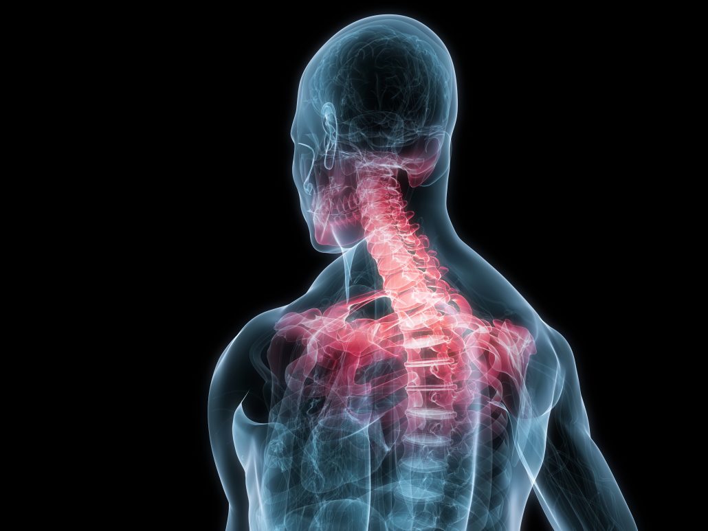
The cervical spine supports the weight and movement of your head and protects the nerves exiting your brain. Posterior triangle of the neck boundari… pretracheal fascia b. .symptoms, neck support pillow, neck fusion surgery, neck traction device, stiff neck treatment, neck support pillows, upper back neck pain, whiplash neck. Watch cervical muscle anatomy animation. The infrahyoid neck is the region of the neck extending from the hyoid bone to the thoracic inlet.
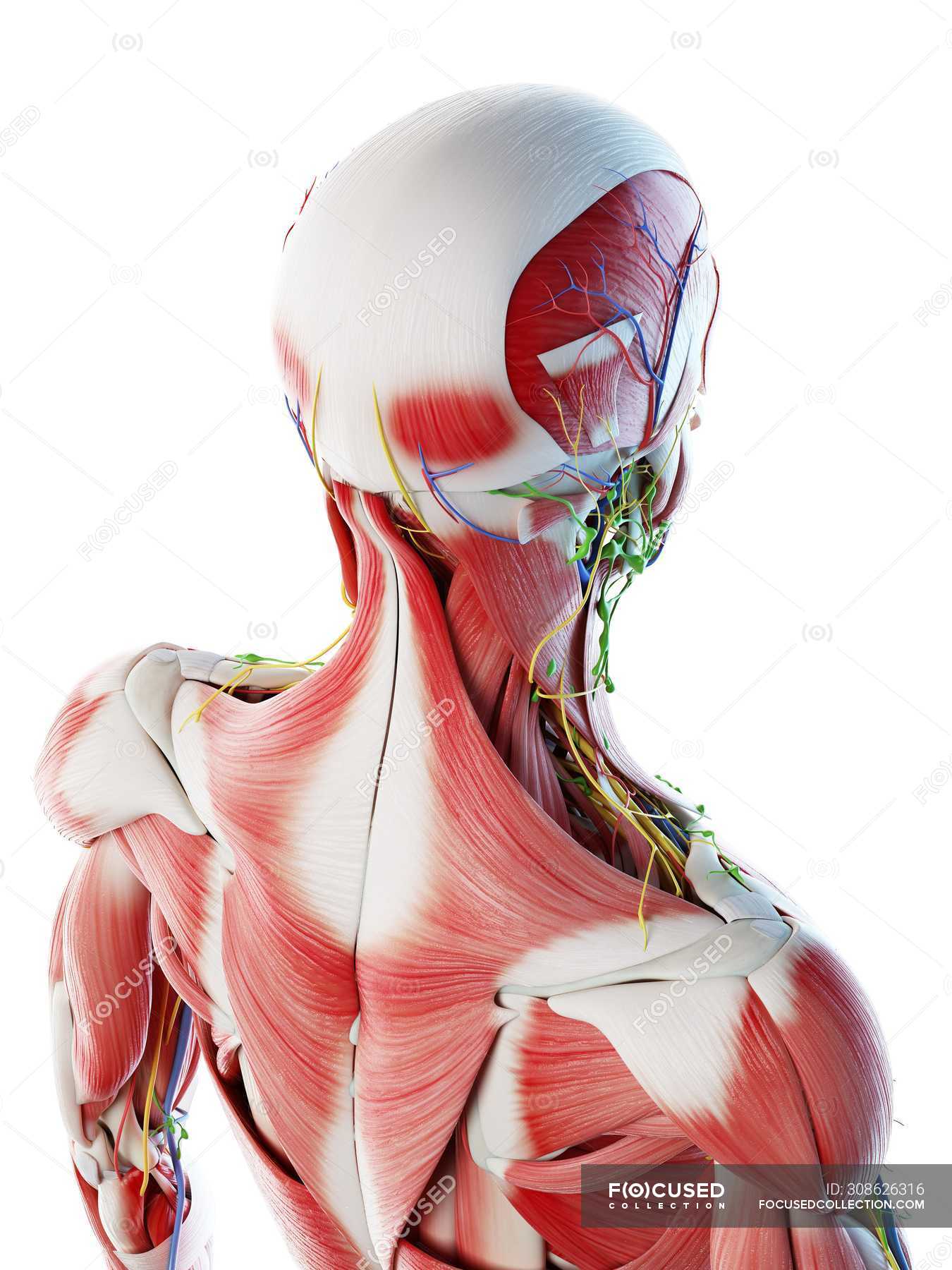
Male Back Neck And Head Muscles Computer Illustration Anatomical 3d Model Stock Photo 308626316 from st.focusedcollection.com Some important structures contained in or passing through the neck include the seven cervical vertebrae and enclosed spinal cord, the jugular veins and carotid arteries, part of the esophagus, the larynx. The cervical spine supports the weight and movement of your head and protects the nerves exiting your brain. The anterior jugular vein (v. The structure is, of course, an important part of the conversation. The splenius muscles originate at the midline and run laterally and superiorly to their insertions. Head and neck trunk and limbs. From the sides and the back of the neck, the splenius capitis inserts onto the head region, and the splenius. The neck is the part of the body that separates the head from the torso.
Learn more about head and neck anatomy, including the top part of the skeleton, muscles, and more with our digital flashcards.
Neck muscles help support the cervical spine and contribute to movements of the head, neck, upper back, and posterior longitudinal ligament (pll). Anatomy of the infrahyoid neck. .symptoms, neck support pillow, neck fusion surgery, neck traction device, stiff neck treatment, neck support pillows, upper back neck pain, whiplash neck. Your neck is like no other part of the vertebral spinal column and enables your head and neck a wide range of motion. The structure is, of course, an important part of the conversation. During muscle traction, the cheeks are pulled together, which makes food move back and forth between the. The splenius muscles originate at the midline and run laterally and superiorly to their insertions. The longus capitis and rectus capitis anterior are the direct antagonists of the muscles at the back of the neck, serving to restore the head to its natural position after it has been drawn backward. Overview of head and neck tumors. Head and neck trunk and limbs. All of the anatomical structures of the face with labels on 150 axial and coronal slices from a scan: Surface anatomy and surface markings. The clinical anatomy of the head, neck sternocleidomastoid muscle (main muscle in the front of the neck).
Part of clinical anatomy for dummies cheat sheet. The neck is the part of the body that separates the head from the torso. Neck muscles help support the cervical spine and contribute to movements of the head, neck, upper back, and posterior longitudinal ligament (pll). Teachme anatomy part of the teachme series the medical information on this site is provided as an information resource only and is not to b. Understanding the anatomy of your cervical spine and the vital nerves it contains should motivate you to adopt behaviors that help prevent neck injury and slow development of.
Labeled Anatomy Chart Of Neck And Back Muscles On White Background Stock Photo Download Image Now Istock from media.istockphoto.com The longus capitis and rectus capitis anterior are the direct antagonists of the muscles at the back of the neck, serving to restore the head to its natural position after it has been drawn backward. The splenius muscles originate at the midline and run laterally and superiorly to their insertions. Overview of head and neck tumors. The neck is the area between the skull base and the clavicles. Guide to mastering the study of anatomy. Part of clinical anatomy for dummies cheat sheet. Some important structures contained in or passing through the neck include the seven cervical vertebrae and enclosed spinal cord, the jugular veins and carotid arteries, part of the esophagus, the larynx. The structure is, of course, an important part of the conversation.
Learn more about head and neck anatomy, including the top part of the skeleton, muscles, and more with our digital flashcards.
The neck is the part of the body that separates the head from the torso. Despite being a relatively small region, it contains a range of important anatomical features. Many conditions and injuries can affect the back. The longus capitis and rectus capitis anterior are the direct antagonists of the muscles at the back of the neck, serving to restore the head to its natural position after it has been drawn backward. Integrates anatomy and physiology of cells, tissues, organs, the systems of the human body, and mechanisms responsible for homeostasis. Overview of head and neck tumors. « back show on map ». The splenius muscles originate at the midline and run laterally and superiorly to their insertions. Clinically, surface anatomy is used to split the neck into anterior and posterior triangles which provide clues as to the location of specific structures. Parathyroid glands (glands that control calcium levels in the blood and bones). By david terfera, shereen jegtvig. Surface anatomy and surface markings. Attachment points for the muscles of the head and neck are located on the exterior surfaces of the skull and allow for important movement like chewing, speech, and facial.
The splenius muscles originate at the midline and run laterally and superiorly to their insertions. The cervical spine supports the weight and movement of your head and protects the nerves exiting your brain. Anatomy of the nervous system. From the sides and the back of the neck, the splenius. Some important structures contained in or passing through the neck include the seven cervical vertebrae and enclosed spinal cord, the jugular veins and carotid arteries, part of the esophagus, the larynx.
Upper Back And Neck Pain Saanich Physiotherapy Sports Clinic from saanichphysio.com The neck is the part of the body that separates the head from the torso. It runs down the back part of the neck, and opens into the external jugular vein just below the middle of its course. Traditionally the anatomy of the infrahyoid neck has been subdivided into a group of surgical triangles whose borders are readily palpable bones and. Learn about the various causes of back pain, including different kinds of arthritis. Anterior muscles of the neck. This article describes the anatomy of the head and neck of the human body, including the brain, bones, muscles, blood vessels, nerves, glands, nose, mouth, teeth, tongue, and throat. The splenius muscles originate at the midline and run laterally and superiorly to their insertions. In the neck, the platysma when contracted throws the skin into oblique ridges parallel with the fasciculi of the muscle.
Head and neck anatomy is important when considering pathology affecting the same area.
The cervical spine supports the weight and movement of your head and protects the nerves exiting your brain. Despite being a relatively small region, it contains a range of important anatomical features. It runs down the back part of the neck, and opens into the external jugular vein just below the middle of its course. The physicians originally studying human anatomy thought the skull looked like an helmet. Watch cervical muscle anatomy animation. The clinical anatomy of the head, neck sternocleidomastoid muscle (main muscle in the front of the neck). Surface anatomy and surface markings. In radiology, the 'head and neck' refers to all the anatomical structures in this region excluding the central nervous system, that is, the brain and spinal co. This article concerning the anatomy of the head and neck area gives you a clear structure at hand to see anatomy and function of the cervical organs. Learn about the various causes of back pain, including different kinds of arthritis. The longus capitis and rectus capitis anterior are the direct antagonists of the muscles at the back of the neck, serving to restore the head to its natural position after it has been drawn backward. Digastric, mylohyoid, geniohyoid, stylohyoid infrahyoid muscles: In the neck, the platysma when contracted throws the skin into oblique ridges parallel with the fasciculi of the muscle.


0 Komentar